
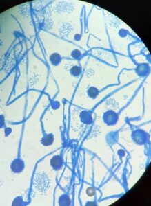
Photo 23
Lactophenol cotton blue (LCB) staining Aspergillus fumigatus
Dr. Harmika Parmar
Photo 22
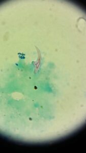
Hooklet of Echinococcus granulosus in Modified ZN Stain preparation from fluid of suspected Hyadatid Cyst of Liver
Dr. Urvesh Shah
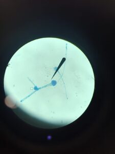
Photo 21
Aspergillus fumigatus in LPCB mount showing conidiophores, philalides covering upper half vesicle and conida
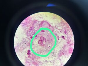
Photo 20
The gram stain from corneal scrap material shows moderate cystic forms of Acanthamoeba species
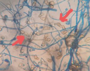
Photo 19
Corneal scrapping – Curvularia spp. Conidia smooth walled, olivaceous brown, obovoidal broadly clavate curved at the subterminal cell, 3 septate and the subterminal cell swollen and distinctly larger than the remaining cells suggestive of Curvularia spp.
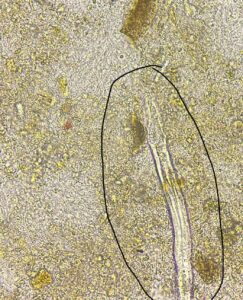
Photo 18
Rhabdatiform larva of Strongyloides stercoralis seen in wet film of stool sample
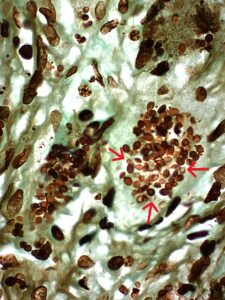
Photo 17
GMS stain of biopsy sample of axillary lymphnode. Small, dark brown to black, oval yeast cells resembling Histoplasma
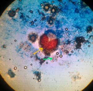
Photo 16
LCB (Lactophenol cotton blue) preparation, Cleistothecium (yellow arrow) of Aspergillus nidulans (anamorph Emericella nidulans) showing numerous reddish-brown ascospores and thick-walled hülle cells (green arrow)
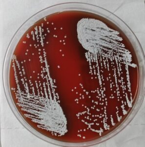
Photo 15
Colonies growth on Sheep Blood Agar showing Nocardia species. It is whitish chalky adherent colonies of Nocardia species. Colonies can be smooth and moist or waxy; with further incubation, development of aerial hyphae causes a velvety or chalky appearance
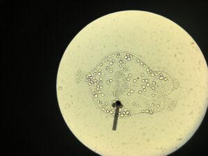
Photo 14
Direct microscopy of the specimen
(Pleural Fluid) is
suggestive of scolex and hooklets of
Echinococcus granulosus
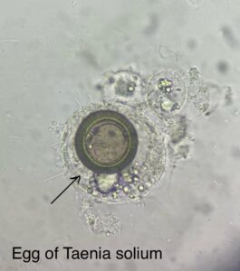
Photo 13
A wet film examination from stool sample showed round eggs consisting of embryo with characteristic six hooklets surrounded by thick radially striated brown shell On ZN stain:
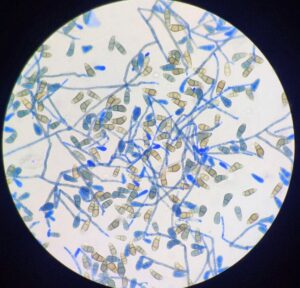
Photo 12
LCB of Curvularia spp. showing septate hyphae with brown colour conidia and central cell of conidia showing swelling which gives curved appearance
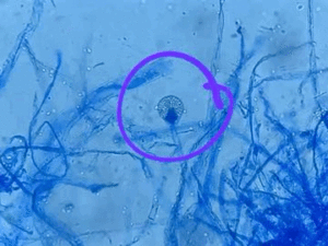
Photo 11
LCB showing Lichtheimia croymbifera fungus in the case of Cutaneous mucormycosis post RTA
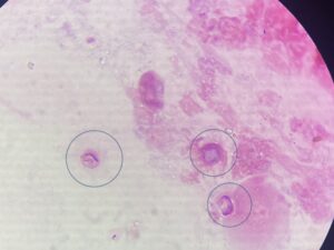
Photo 10
Gram stain from corneal scraping showing cysts of Acanthamoeba
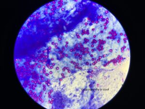
Photo 9
Modified ZN stain of stool sample showing acid fast, round to oval, structures suggestive of oocysts of Cryptosporidium species
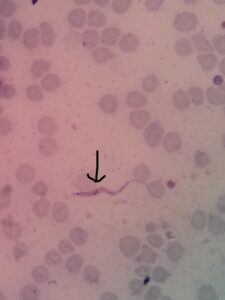
Photo 8
Peripheral smear showing flagellated parasite with subterminal kinetoplast resembling trypanosomiasis
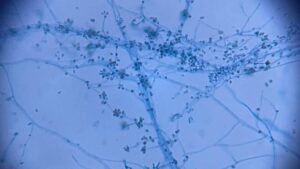
Photo 7
Hyaline, thin septate hyphae with globose conidia
Stain: LPCB (lactophenol Cotton Blue) stain
Fungi: Sporothrix globosa
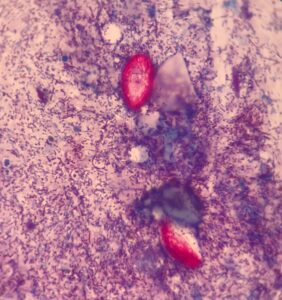
Photo 6
Sample: Stool
Oocysts of Cystoisospora belli stained with the Modified acid-fast stain
Photo 5
T. Cruzei
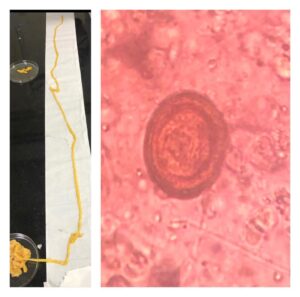
Photo 4
Left side image shows Taenia Saginata worms which are approx 5-6 meters in length.
Right side image shows Taeina Saginata eggs in stool. The eggs are spherical and brown in colour .Size of eggs approx 30-35 micrometers in diameter and radially striated. The internal Onco sphere contains refractile hooklets.
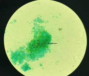
Photo 3
Specimen- Bronchoalveolar lavage (BAL)
GMS (Gomori Methenemine silver) preparation-
Cyst wall is stained black, organism appear as folded & flattened spheres characteristic of Pneumocystis jirovecii (carinii)
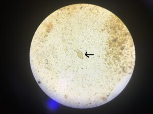
Photo 2
Eggs of Fasciola hepatica in duodenal aspirate. Large (140-180 µm), operculated, Ovoid in shape, bile- stained. Contains a large unsegmented ovum.
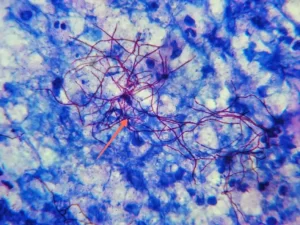
Photo 1
Modified acid fast staining using 1% sulfuric acid as decolorizer (Kinyoun method) shows partially acid-fast branching and filamentous red colored acid-fast bacilli suggestive of Nocardia species.
CONTACT US FOR MORE INFORMATION |